
Photoacoustic Tomography for Human Brain
Blood-oxygen-level-dependent (BOLD) functional magnetic resonance imaging of the human brain requires bulky equipment for the generation of magnetic fields. Photoacoustic computed tomography obviates the need for magnetic fields by using light and sound to measure deoxyhaemoglobin and oxyhaemoglobin concentrations to then quantify oxygen saturation and blood volumes. Here, we show that massively parallel ultrasonic transducers arranged hemispherically around the human head can produce tomographic images of the brain with a 10-cm-diameter FOV and spatial and temporal resolutions of 350 µm and 2 s, respectively. A comparison of functional photoacoustic computed tomography and 7 T BOLD functional magnetic resonance imaging showed a strong spatial correspondence in the same FOV and a high temporal correlation between BOLD signals and photoacoustic signals, with the latter enabling faster detection of functional activation. Our findings establish the use of photoacoustic computed tomography for human brain imaging.
Empowered with a high spatiotemporal resolution and sensitivity, the 1K3D-fPACT can image both brain function and angiographic structures, facilitating coregistration between the fPACT and fMRI results. The 1K3D-fPACT angiography and MRA result in a strong visuospatial correlation. Quantitively, the measured vasculature diameters of participant 1 exhibit a strong agreement. The potential of the 1K3D-fPACT to diagnose cerebral vascular diseases is evident.
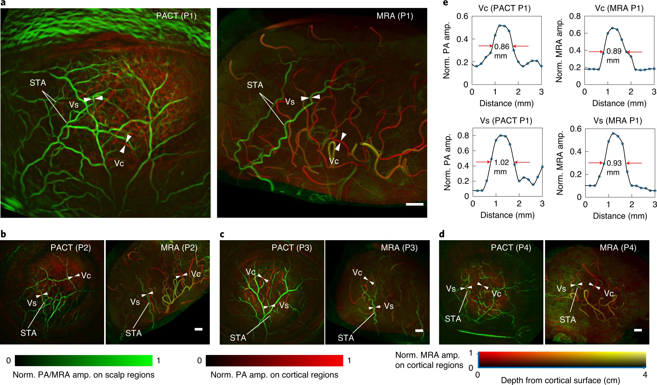
Comparisons of the angiographic structures and functional maps acquired on 7 T MRI and the 1K3D-fPACT for participant 1.
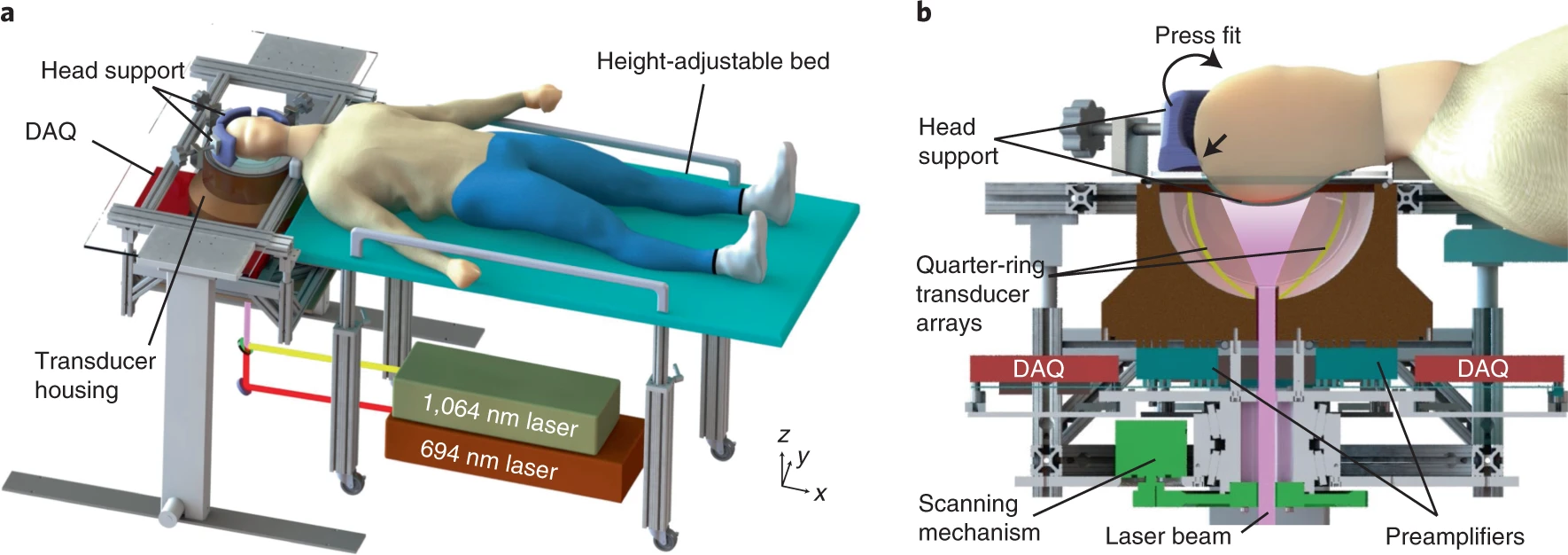
Participant 1 being imaged by the 1K3D-fPACT when performing the finger-tapping task.
Publications and links
- Massively parallel functional photoacoustic computed tomography of the human brain, Nature Biomedical Engineering 2022.
- High-speed three-dimensional photoacoustic computed tomography for preclinical research and clinical translation](https://ieeexplore.ieee.org/abstract/document/7951481), Nature communications 2021.
- Transcranial photoacoustic computed tomography based on a layered back-projection method, Photoacoustics 2020.
- Real-time tomography of the human brain
- 光声成像新突破:人脑功能层析成像